
A better evaluation of liver fibrosis
Shear wave elastography (SWE) offers better early assessment

Jane Sohn, MD
Liver disease is an important problem with an estimated worldwide mortality of 1.5 million per year. It is the 12th leading cause of death in the US and represents a significant burden to healthcare providers.
Liver injury arises from a wide range of causes, including viral hepatitis, alcoholic and nonalcoholic fatty liver disease, drug-induced liver injury, primary biliary cirrhosis, and autoimmune hepatitis. Medications and obesity also can cause excessive accumulation of extracellular matrix proteins within the liver parenchyma leading to fibrosis and eventually cirrhosis.
Accurately staging liver fibrosis is essential for therapeutic decisions, prognostic evaluation and ongoing management and the need is growing for simple, repeatable, and cost-effective diagnostic tools.
Limitations of liver biopsy for fibrosis
While liver biopsy and histological assessment of fibrosis is regarded as the “gold standard”, biopsy remains a relatively inefficient determination of fibrosis. Even optimally sized liver biopsies reflect a tiny fraction of the organ, which is insufficient in diseases where fibrosis can be unevenly distributed, and additional sampling increases risk of complications. Consequently, sampling error may occur in up to 25-30% of liver biopsies.1
Given these limitations of biopsy, and patients’ desire to avoid invasive testing, researchers have done much work over the past 10 years to develop noninvasive tests that can measure liver fibrosis.
Shear wave elastography (SWE) vs transient elastrography (TE) for fibrosis
Transient elastography with FibroScan® first became available 13 years ago. It tracks the velocity of shear waves generated by the gentle hit of a piston on the skin, with the resulting compression wave traveling in the liver along its longitudinal axis. TE provides quantitative information about the stiffness of the liver and hence an assessment of the amount of fibrosis in chronic liver disease. It is a noninvasive test that has not been associated with any side effects.
Jane Sohn, MD Head of Ultrasound Imaging at SMIL says a more precise method of shear wave elastography (SWE) is now available at Scottsdale Medical Imaging (SMIL) for the evaluation of liver fibrosis.
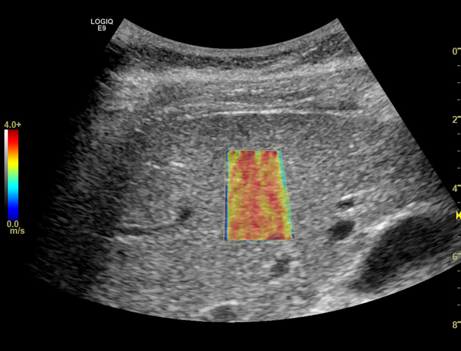
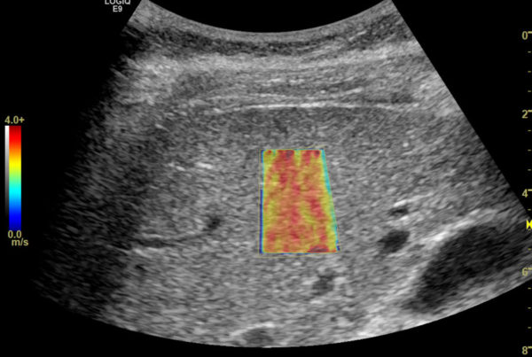
Unlike TE, SWE enables the measurement of liver stiffness where desired, by watching a real-time image with B-mode ultrasound, In addition, SWE can assess the homogeneity of the liver, because SWE produces color images corresponding to the varying degrees of stiffness. Due to these advantages, SWE has been suggested as a more precise method of the measurement of liver stiffness than TE.2
“SWE makes it possible to use B‐mode ultrasound images to specifically identify the region within the liver from which measurements can be obtained,” Dr. Sohn says. “An additional advantage is B‐mode ultrasound interrogation of the liver, spleen and vascular anatomy can also be performed during a single appointment for the patient and rather than needing different transducers SWE uses a single transducer with dual frequencies that can be manipulated dependent on patient body habitus.”
In a comparison of TE and SWE Ferraioli and colleagues found SWE to be more accurate than TE in assessing significant fibrosis (F2 and greater).3 In addition the researchers found SWE has the advantage of imaging liver stiffness in real time while guided by a B-mode image. Thus, the region of measurement can be guided with both anatomical and tissue stiffness information.
Other studies have shown SW elastography enables a better biopsy-correlated assessment of liver fibrosis compared with transient elastography, especially for early fibrosis. SWE elastography also gives objective assessment of splenic elasticity, correlated with liver fibrosis staging in patients with chronic hepatitis B.
Given the research evidence, Dr. Sohn says SMIL believes SWE offers referring physicians superior diagnostic confidence compared with the former standard of liver biopsy and better overall evaluation than transient elastography, providing patients better early assessment of liver fibrosis.
__________________
1F.M. Sanai and E.B. Keeffe; Liver Biopsy for Histological Assessment –The case against. Saudi J Gastroenterol. 2010 Apr; 16(2): 124–132.
2Bavu E, Gennisson JL, Couade M, et al. : Noninvasive in vivo liver fibrosis evaluation using supersonic shear imaging: a clinical study on 113 hepatitis C virus patients. Ultrasound Med Biol, 2011,37: 1361–1373. [PubMed]
3Ferraioli G, Tinelli C, Dal Bello B, et al.: Accuracy of real-time shear wave elastography for assessing liver fibrosis in chronic hepatitis C: a pilot study. Hepatology. 2012 Dec;56(6):2125-33