
Identifying Risk of Osteoporotic Fracture
DXA combined with FRAX provides more accurate assessment

Jimmy Leung, MD
The National Osteoporosis Foundation estimates that 10 million Americans are affected by osteoporosis and roughly 34 million more are at risk for the disease because of low bone density.
“The problem with osteoporosis, much like breast cancer, is that it can be a completely asymptomatic disease until a fracture occurs,” says Jimmy C.M. Leung, M.D., a musculoskeletal radiologist at Scottsdale Medical Imaging Ltd. (SMIL). “And like breast cancer, it can be a source of mortality and is a high source of morbidity for both men and women.”
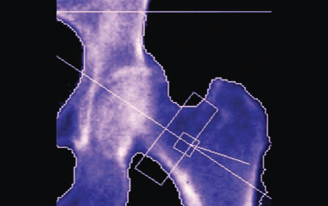
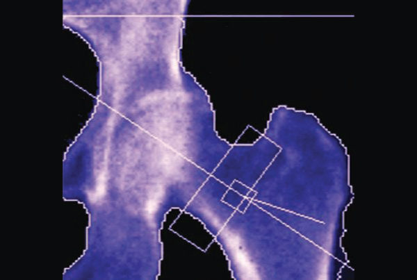
Dual X-ray absorptiometry (DXA) is the primary screening method to detect the disease. Using a fraction of the radiation dose used for a chest X-ray, a DXA scan provides a T score of the patient’s bone mineral density compared with a population-wide database of peak bone mass calculated to reflect the person’s age.
Combining DXA with the World Health Organization’s Fracture Risk Assessment Tool (FRAX®) to adjust for individual characteristics, clinicians can gain even more confidence in determining pharmacological treatments. Applying the FRAX tool, however, can be time-consuming.
“At SMIL, we now incorporate the FRAX tool in our reports,” Leung says. “We’ve always been doing densitometry to estimate bone loss. Adding FRAX produces an absolute risk. It can tell you that people with this bone density and this demographic data have this much risk in the next 10 years of having a fracture.” •