
Imaging Options for Acute Stroke and TIA Patients
Unraveling the complexity of evaluation techniques

Steven Wise, MD
A wide variety of imaging techniques has become available to assess vascular lesions and brain tissue status in acute stroke patients. As a result of the multiple facets of these imaging techniques and complex algorithms, evaluation of patients for acute stroke or transient ischemic attack (TIA) is more complex than ever.
While the complexity of techniques is new, Steven Wise, MD, a neuroradiologist at Scottsdale Medical Imaging (SMIL), says that in the emergency setting some things remain the same.
“If the patient is within the zero- to six-hour window of an acute stroke, we want to make sure they are sent to the emergency department, not to the outpatient SMIL facility,” Wise says. “A noncontrast CT of the head should be obtained to determine eligibility for IV tPA or emergent neuroendovascular intervention, which could prevent a major stroke.”
Outside that six-hour window, or when initial stroke evaluation is inconclusive, Wise says that further workup of patients with neurologic deficits may benefit from MRI brain and MR angiography (MRA) of the head and neck.
“We’re introducing a new stroke/TIA protocol that includes MRI brain with diffusion weighted imaging and MRA of the head and neck, all without contrast,” Wise says. “One reason is that MRI brain is far more sensitive for detecting small infarcts than CTA and CT perfusion. Some of the elderly members of our community will often have these smaller infarcts, which will likely not be detected on other techniques.”
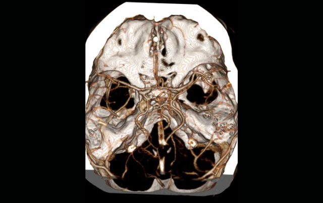
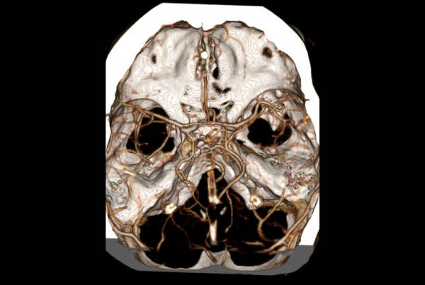
For MRA of the head, Wise says, SMIL radiologists will evaluate for a flow-limiting stenosis or an occluded major vessel. For the neck, they look for carotid and vertebral artery disease. In patients with neck pain, they might add a dissection protocol to the MRA of the neck, especially if it is a young patient having a stroke.
MRA neck can be performed with or without IV contrast if the proximal great vessels need to be evaluated, such as in patients with subclavian steal or vertebrobasilar symptoms.
CTA of the head and neck, either without or with CT perfusion with IV contrast is the imaging workup of choice in patients who are not eligible for MRI. •
REFERENCE:
Wintermark M, Sanelli P, Albers G, et al. Imaging recommendations for acute stroke and transient
ischemic attack patients: a joint statement by the American Society of Neuroradiology, the American
College of Radiology, and the Society of NeuroInterventional Surgery. AJNR Am J Neuroradiol.
2013;34:E117–E27.
