
New Options for Imaging in Crohn’s Disease
Combining CT and MRE effective for assessing small bowel disease

Mark Kuo, MD
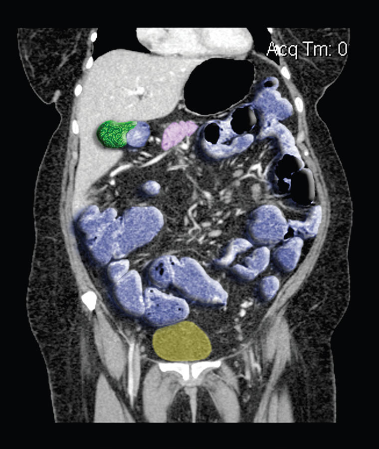
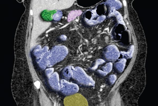
Until recently, the gold standard for evaluating small bowel disease, including Crohn’s disease, has been enteroclysis. This is gradually being replaced by less invasive CT and MR enterography (MRE) due to improved spatial and temporal resolution combined with improved bowel distending agents.
In general, patients will find MRE less invasive compared with enteroclysis, says Mark Kuo, MD, a radiologist at Scottsdale Medical Imaging Ltd. (SMIL).
“They do have to drink a little bit of contrast material before the study,” Kuo says. “What that does is distend the small bowel so we can see it better. Then they get on the MR table and they undergo several sequences, and one of them includes an IV contrast administration.”
While concerns about ionizing radiation exposure from CT for chronic diseases have slowed widespread adoption of this technology, Kuo says SMIL protocols address this concern by combining CT with MRE.
“Because patients with chronic ailments, such as Crohn’s disease, often need repeat imaging, CT radiation dose can accumulate over time,” Kuo says. “This is especially true for many Crohn’s patients, who are diagnosed at a young age. In order to minimize radiation dose, SMIL utilizes MR enterography, which has no associated radiation.”
He says SMIL enterography protocols use CT for initial diagnosis and MRE for follow-up evaluations. Together the results provide high-quality imaging with substantially reduced radiation exposure.
“Typically, we like to have one baseline CT because there is a little less motion on a CT than with MRE,” Kuo says. “After that, we can do MRE with Crohn’s patients who are having recurrence of abdominal pain instead of having to undergo multiple CT scans with associated radiation. We can use MRE to find out if there is active disease, where it is and characterize it without having to undergo another CT scan.”
Kuo notes that MRE does have limitations for some patients, including those with a pacemaker or an aneurysm. •
REFERENCE:
Siddiki Hassan, Fidler Jeff. MR imaging of the small bowel in Crohn’s disease. European Journal of
Radiology. 2009; 69(3):409-417.
